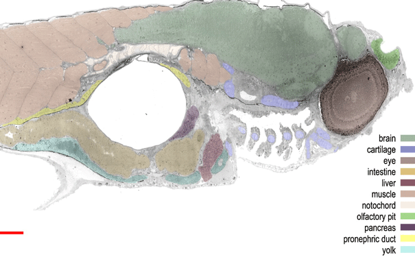
Virtual nanoscopy: High resolution image of a zebra fish embryo sagittal section. Image: Williams et al 2012, JCB
Advances in microscopy allow researchers to zoom in and out of biological tissue like they’re using Google Earth.
Tissue imaging techniques range from simple cell stains to advanced electron microscopy. A great deal of information on cellular structure and physiology has been revealed from being able to observe biological tissue at a nanometre resolution.
Existing visualisation techniques have their limits, however, in that they can only capture a small chunk of the cell in a single snapshot. This means researchers are unable to determine how the structures they see relate to other areas of the cell and to the organism as a whole.
A team of researchers from Leiden University Medical Centre in the Netherlands has developed a new technique called virtual nanoscopy, which will resolve the problem of having only a small field of view in a single image. They stitched together over 26,000 images to generate an almost complete ultrastructural map of a zebrafish embryo. The large-scale micrograph had a resolution of 16 million pixels per inch (2.54 centimetres) and contained 281 gigapixels.
“[This technique] will change the way electron microscopy is done; instead of collecting just a snapshot of part of a cell, we now routinely collect the entire cross-section of that cell which provides a much better context of the observed phenomena,” says Raimond Ravelli, co-author of the study published in the Journal of Cell Biology (JCB).
Just as one can explore the world, countries and streets on Google Earth, researchers will now be able to navigate through biological tissue from the level of the whole organism down to its subcellular structures. The zebrafish image from the current study, among others, is publicly accessible to explore using a program called JCB DataViewer.
“The JCB DataViewer was launched in 2008 to promote sharing of original data,” explained Elizabeth Williams, JCB executive editor. “If you can image it, you should be able to publish it.”
“For the future, we are optimistic that hardware improvements (such as a new generation of much faster detectors) as well as automated segmentation procedures might revolutionise cell biology by high-resolution 3D analysis of cells and tissues at different stages of disease,” says Ravelli.
Source: Eurekalert